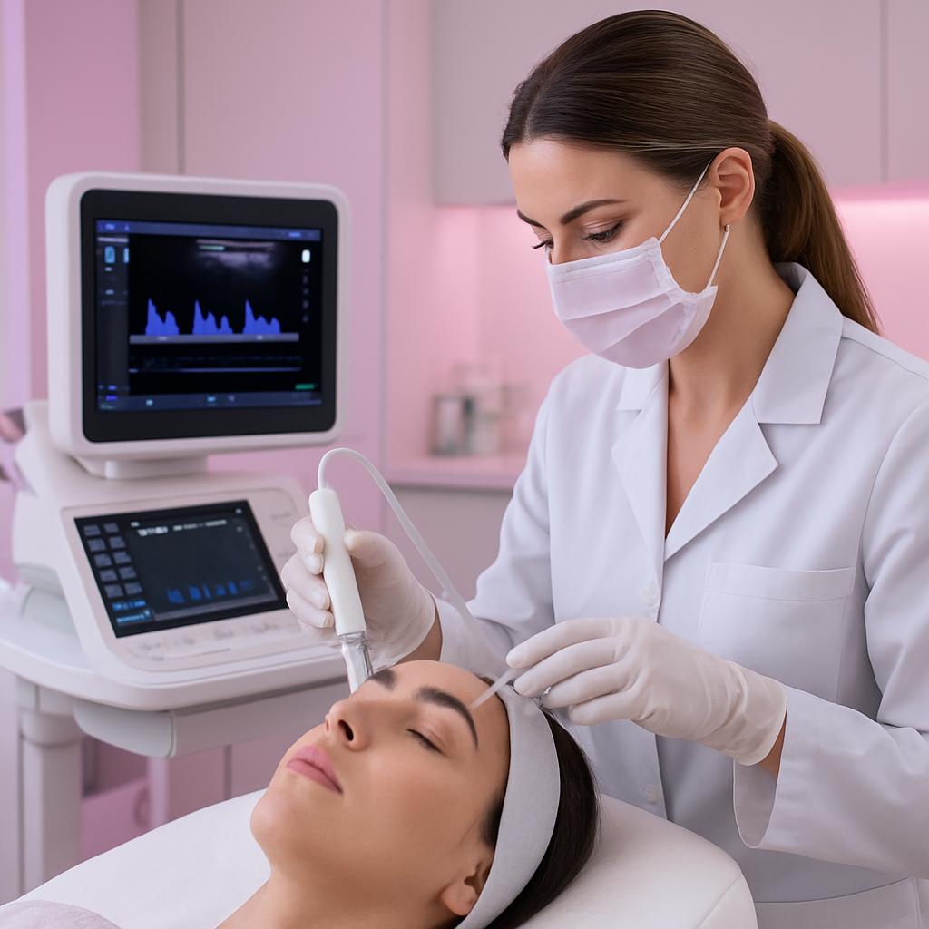
While anatomical mapping provides essential knowledge of facial structures, ultrasound Doppler technology takes safety to the next level by allowing us to see blood vessels in real-time. At Magenta Clinique, we combine expert anatomical knowledge with advanced imaging for maximum patient safety.
This technology is not required for every treatment, but for high-risk areas or complex cases, ultrasound guidance provides invaluable visual confirmation that was previously impossible. Learn how this cutting-edge approach is changing the safety profile of filler injections.
What is Ultrasound Doppler Technology?
Seeing Beyond the Surface
Ultrasound uses high-frequency sound waves to create images of structures beneath the skin. Doppler mode adds the ability to detect movement specifically blood flow allowing us to see exactly where arteries and veins are located. Color Doppler displays blood flow direction, with arteries and veins showing in different colors.
B-Mode Ultrasound
Grayscale imaging showing tissue layers, fat, muscle, and structures like blood vessels as dark circular areas.
Uses: Identifying tissue planes, locating old filler, measuring depth
Color Doppler Mode
Adds color to show blood flow within vessels. Red typically indicates flow toward the probe, blue indicates flow away.
Uses: Confirming vessel location, distinguishing arteries from veins
High-frequency probes (15-22 MHz) used for aesthetic procedures provide excellent resolution for superficial facial structures. This allows visualization of even small blood vessels just millimeters below the skin surface.
Why Vascular Safety Matters in Filler Treatment
Understanding the Risk
Vascular occlusion occurs when filler enters or compresses a blood vessel, blocking blood flow. The face contains vessels that connect to the ophthalmic artery (which supplies the eye) and intracranial circulation. While rare, vascular events can lead to:
- •Skin necrosis (tissue death) in the affected area
- •Vision changes or, in severe cases, vision loss
- •Stroke-like symptoms (extremely rare)
According to research published in Aesthetic Surgery Journal, the use of ultrasound in aesthetic procedures is increasingly recognized as a valuable safety tool, particularly for high-risk injection sites.
Prevention is always better than treatment. By seeing blood vessels before we inject, we can avoid them entirely rather than relying solely on technique and anatomical knowledge.
How Ultrasound-Guided Filler Injection Works
Pre-Treatment Scanning
Before any injection, the treatment area is scanned to identify blood vessel locations, depths, and any anatomical variations. This creates a vascular map specific to your face.
Vessel Marking
Key vessels can be marked on the skin surface to create visual guides during injection. This complements anatomical landmarks.
Real-Time Guidance (When Indicated)
For the highest-risk areas, the ultrasound probe can be held adjacent to the injection site, providing real-time visualization during filler placement.
Post-Injection Verification
After injection, ultrasound can verify proper filler placement and confirm no vascular compromise has occurred.
When is Ultrasound Guidance Recommended?
Not every filler treatment requires ultrasound. However, it adds significant value in specific situations:
Temples
Why: Superficial temporal artery and its branches are clearly visible on ultrasound
Benefit: Direct visualization allows safe navigation around critical vessels
Tear Troughs
Why: Angular artery and proximity to orbit make this a delicate area
Benefit: Confirms proper depth and avoids vascular structures
Nose (Non-Surgical Rhinoplasty)
Why: Dorsal nasal artery with ophthalmic artery connections
Benefit: Maps vessel location before any product placement
Filler Correction Cases
Why: Old filler may have migrated or be in unexpected locations
Benefit: Locates filler deposits for targeted dissolution with hyaluronidase
Revision Patients
Why: Previous treatments may have altered normal anatomy
Benefit: Provides clarity before treating patients with complex history
Benefits of Ultrasound Doppler Guidance
Real-Time Visualization
See blood vessels on screen as you inject, not relying solely on anatomical knowledge
Dynamic Assessment
Observe how tissues and vessels respond during injection in real-time
Identify Old Filler
Locate previous filler deposits for precise correction or avoidance
Enhanced Confidence
Greater certainty in high-risk areas where anatomical variation is common
Beyond Safety: Additional Applications
- Locating old filler: For correction treatments, ultrasound can identify exactly where previous filler sits
- Monitoring complications: If vascular compromise is suspected, ultrasound can assess blood flow
- Guiding dissolution: Targeted hyaluronidase placement for precise correction
- Treatment planning: Assess tissue quality and volume for optimal approach
Our Approach at Magenta Clinique
Integrated Safety Protocol
At Magenta Clinique, ultrasound Doppler is one component of our comprehensive safety approach:
- Foundation of anatomical knowledge: Dr. Maggie Fawzi has extensive training in facial anatomy and anatomical mapping
- Appropriate technique selection: Cannula vs. needle based on area and risk
- Ultrasound when beneficial: Not used unnecessarily, but available when it adds meaningful safety
- Emergency preparedness: Hyaluronidase and protocols always ready
We believe in using technology thoughtfully not as a marketing gimmick, but as a genuine tool for enhanced patient safety. Dr. Fawzi will discuss whether ultrasound guidance is recommended for your specific treatment plan.
Frequently Asked Questions
Is the ultrasound procedure painful?
Not at all. Ultrasound scanning is completely non-invasive and painless. A small amount of gel is applied to the skin, and the probe is moved gently over the treatment area.
How long does ultrasound assessment add to treatment time?
Pre-treatment scanning typically adds only 5-10 minutes. For real-time guided injection, treatment time may be slightly longer, but the safety benefit far outweighs this small time investment.
If ultrasound is available, why is not it used for everyone?
For many treatment areas (like lips or superficial cheek augmentation), proper anatomical knowledge and technique provide adequate safety. Ultrasound adds most value in high-risk areas. We recommend it when the benefit justifies the added time and cost.
Can ultrasound detect all blood vessels?
High-frequency ultrasound can detect most clinically significant vessels in superficial facial tissues. Very small capillaries may not be visible, but these are not typically associated with serious complications. The major arteries of concern are reliably visualized.
Experience the Safest Filler Treatment in Cairo
Book a consultation with Dr. Maggie Fawzi at Magenta Clinique. We will discuss whether ultrasound-guided injection is right for your treatment goals.
Magenta Clinique, Maadi, Cairo